The future of medical modeling

The Problem

Creating patient-specific 3D models is time consuming, difficult, and tedious.

The Reason

Automation can only help so much since current platforms are 2D tools trying to perform 3D tasks.

The Solution

We’ve designed Elucis, the world’s first VR platform for creating and using 3D medical models. For the first time, you can create 3D medical content in 3D!

How it works at a glance

A 3D CT scan intersecting a visualization plane in VR
A VR stylus drawing on a medical image and creating a 2D model
A VR stylus drawing on a 3D medical model with a CT image

Learn more about how Elucis is different

Watch the videos below to learn about all of Elucis' exciting and powerful features.

Your medical images exist in the environment as real 3D volumes that can be fluidly and intuitively reoriented. In combination with one or more viewing planes, any conventional or arbitrary multiplanar reformation can be quickly accessed and understood.  A 3D ghost view of the image is automatically displayed when repositioning the image volume to provide meaningful context to the image’s orientation.  Elucis unlocks the use of thin-slice image data in a powerful and meaningful way.

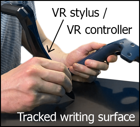
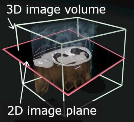
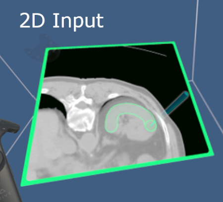
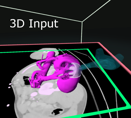
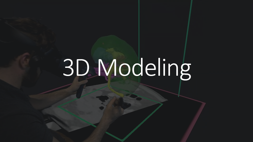
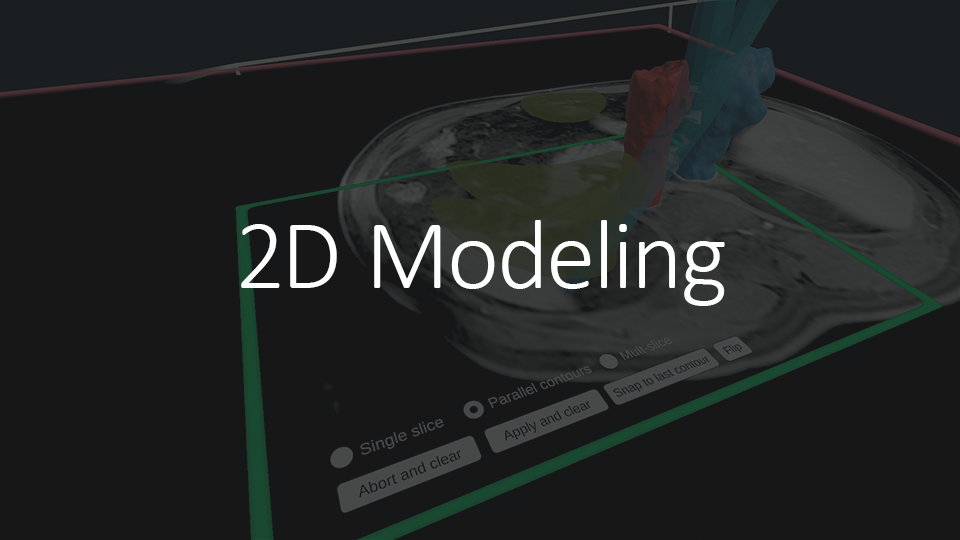
Anatomical structures can be created by drawing directly on these planar image views using a virtual reality stylus that can been seen in VR.  Or, create and edit structures by drawing on the image off of the viewing plane and in mid-air.  Since our modeling tools operate in 3D, there’s no difference between planar or mid-air creation. One simply decides how to create at any given moment based on the need to refer to planar views, or to judge the 3D view of structures.

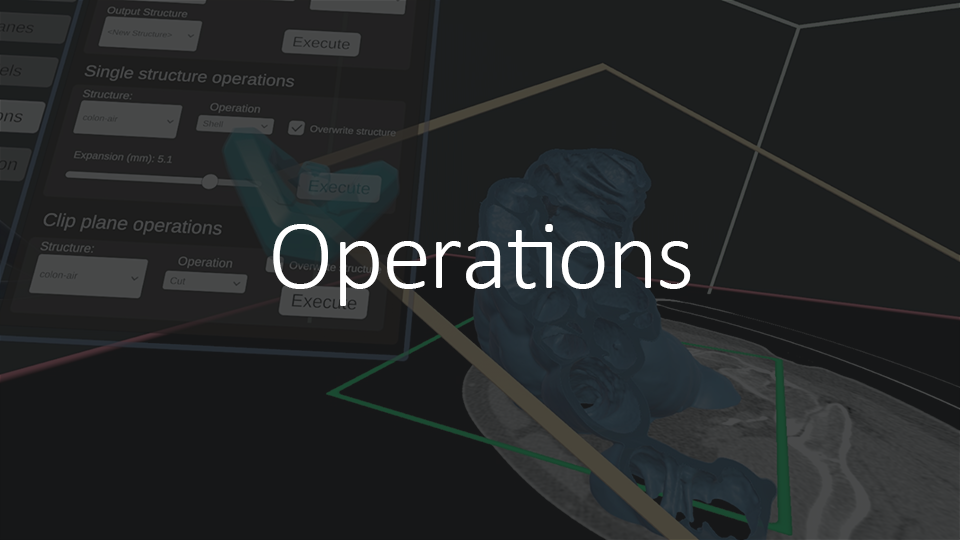
Elucis uses intelligent 2D and 3D operations to quickly model regions of interest based on user input, image values, and a few simple, user-defined settings. 3D structures of your patient’s anatomy appear in real time and you have full control over its visual characteristics, including the size,  color, and transparency.

We don’t use the term “segmentation” to refer to our modeling activities. That is because it’s never just segmentation, but the full end-to-end process in one step. The moment you start drawing, you have a fully finished and usable model in front of your eyes, ready to be exported as an STL or OBJ file. 2D and 3D views always represent the fully realized 3D structures. That means that in the images you are seeing where structures seem to be appearing with a wave of hand, there are no additional steps needed to have high resolution export-ready meshes. To that end, Elucis is the perfect precursor to medical 3D printing. Structures created in Elucis are “water-tight” and can be further post-processed for 3D printing. Alternatively, import models into other applications of your choice for use digitally. They are even compatible with radiation oncology treatment planning system software.

Since structures are never just segmentations, an array of 3D post-processing operations such as Boolean operations, expansions, duplications, global smoothing, shell operations, or fluidly previewing and applying cutting planes are available at any point in the modeling process.

While Elucis features intuitive 3D modeling tools that allow you to create 3D models from thresholded volumes-of-interest with full control, a range of 2D editing tools is also available.

Users have the option of importing multiple fused images, such as a co-registered CT and MRI scans, so they can use the image that best reveals the anatomy of interest for modelling. Seamlessly switch between the two images, or use a blended view. Image thresholding and window and level settings are set independently.

Elucis sports a sleek desktop interface that allows users and collaborators to:

  • quickly import or export images and models, or load existing projects,
  • define custom settings,
  • review 2D cross-sections,
  • leave annotations,
  • explore tutorials, and more.

Elucis has a powerful 3D selection function that can be used for a number of operations. Multiple selection tools can be used to quickly highlight 3D regions of interest.  Users can display a preview of their selection and can modify the selection on the fly during this preview visualization.  Selections can be used to delete,  copy, or split structures.

In addition to being a powerful model creation tool, Elucis can also import 3D structures created in other platforms in the form of OBJ, STL, and Nifty files as well as radiotherapy contours. Imported structures are quickly converted into Elucis’ internal format and can be edited just like any other structure created from within the platform.
Imported structures and structures created from scratch can also be unlocked to become grabbable objects that can be repositioned or resized.

Elucis has a number of asynchronous and synchronous communication tools.  Users can quickly create 3D measurements in the scene, or easily markup structures with 3D sketches to add communicative context to structures.  Additionally, users of the desktop user interface can point out parts of images and structures by drawing with the mouse on the conventional image viewing panels.  The user in the VR environment will see where they’re pointing with a glowing ball at that mouse cursor’s position.  Users can also communicate by adding voice memos that are associated with landmarks or in 3D space.  Voice memos are a powerful tool for asynchronous collaborative model creation workflows as well as for communicating details to downstream recipients of your 3D content.  They can be created or played back in the 3D virtual environment, or from the conventional image viewing panels in the desktop user interface.

Elucis has fully integrated remote networked collaboration. Connect with other users in a shared virtual environment that allows full simultaneous structure creation and editing. The images, 3D structures, and all patient identifying information stays entirely within your custody at all times. In-house remote collaborations can be facilitated with a single click by storing a project database on a network drive or institutionally approved cloud storage drive of one’s choice. If users want to collaborate externally, they can do so by exporting an Elucis project and sharing it with their collaborator through secure means of their choosing. A remote session can then be created on the fly through the generation of a secure one-time key. Additionally, the telemetry facilitating the remote interactions never contains image or 3D structure data, and voice communication is always encrypted.

Supported data formats

Input

    • DICOM CT
    • DICOM MRI

Model import into editable format

    • NifTi images and models
    • STL
    • OBJ
    • DICOM RT Structure

Output

  • STL
  • OBJ
  • DICOM RT Structure

Coming soon:

  • NifTi models
  • DICOM encapsulated STL & OBJ

System Requirements

Elucis is a virtual reality-based platform that runs on SteamVR

Compatible VR systems:

  • SteamVR Tracking-based VR systems such as Vive, Vive Pro, Valve Index
  • PC-based Oculus VR systems + Oculus Quest (with link cable)
  • Compatible with all VR controllers

Recommended minimum system specifications:

  • NVIDIA GTX 1080 or higher
  • Intel Core i7-6700K
  • 32 GB RAM (the more the better)
Illustration of how to set up working space for VR based medical modeling with Elucis

Interested in trying the platform?

Request your free 30-day trial today!